|
Overview
The condition of flat feet in adults is known as ?fallen arches.? Not all adults develop flat feet, and some people are more prone to developing the condition than others. An obese person puts extra weight on their feet while walking or standing. Over time, this can weaken the components that make up the arch and cause the arch to collapse. A woman who is pregnant may also suffer from flat feet during her pregnancy. The problem with developing flat feet as an adult is that in most cases the changes are permanent, if not bothersome. Doctors recommend using custom-made orthotics in shoes to treat the problem. Flat feet were once considered a result of poor health, but it has been proven that athletes such as runners, who are in great condition, also suffer from flat feet. In fact, it?s very common among track runners. Flat feet were once thought of as a bad thing. But studies show that people with higher arches are four times more likely to injure or sprain their ankles than people with flat feet. Studies conducted by the military have discredited the idea that flat feet are a reason to be excused from service. Causes Family history, experts say fallen arches can run in families. Weak arch, the arch of the foot may be there when no weight is placed on it, for example, when the person is sitting. But as soon as they stand up the foot flattens (falls) onto the ground. Injury, arthritis, tibialis posterior (ruptured tendon), pregnancy, nervous system or muscle diseases, such as cerebral palsy, muscular dystrophy, or spina bifida. Tarsal Coalition, the bones of the foot fuse together in an unusual way, resulting in stiff and flat feet. Most commonly diagnosed during childhood. Diabetes. Age and wear and tear, years of using your feet to walk, run, and jump eventually may take its toll. One of the eventual consequences could be fallen arches. The posterior tibial tendon may become weakened after long-term wear a tear. The postario tibial tendon is the main support structure of the arch of our feet. The tendon can become inflamed (tendinitis) after overuse - sometimes it can even become torn. Once the tendon is damaged, the arch shape of the foot may flatten. Symptoms Most people do not exhibit any symptoms of flat feet, but if the condition is due to an underlying problem, symptoms can include foot pain, mainly in the arch or heel areas, difficulty standing on tiptoes, swelling that occurs on the inside of the ankle, pain in the calf, knee, hip, or lower leg area, both feet lie flat on the ground with no gap, Shoes may not fit properly, heel may tilt away from the midline of the body more than usual, absence of foot arch when standing. If you are experiencing these symptoms and have flat feet, you should consider seeing your doctor or a podiatrist immediately for an examination. Diagnosis Your doctor examines your feet to determine two things, whether you have flat feet and the cause or causes. An exam may include the following steps, Checking your health history for evidence of illnesses or injuries that could be linked to flat feet or fallen arches, Looking at the soles of your shoes for unusual wear patterns, Observing the feet and legs as you stand and do simple movements, such as raising up on your toes, Testing the strength of muscles and tendons, including other tendons in the feet and legs, such as the Achilles tendon or the posterior tibial tendon, Taking X-rays or an MRI of your feet. deelsonheels Non Surgical Treatment If your condition is bothersome, try elevating your feet and using ice on the arches to reduce swelling. Your podiatrist can recommend several orthotic aids and inserts to strengthen the tendons of your foot. He can also demonstrate stretching exercises or refer you to physical therapy to get those tendons back into shape. If the symptoms of fallen arches are painful and troubling, he may recommend a steroid injection to relieve inflammation and pain. And in some instances, he may determine that surgery is necessary. Surgical Treatment  Common indications for surgery are cerebral palsy with an equinovalgus foot, to prevent progression and breakdown of the midfoot. Rigid and painful Pes Planus. To prevent progression, eg with a Charcot joint. Tibialis posterior dysfunction, where non-surgical treatment is unsuccessful. Possible surgical procedures include Achilles tendon lengthening. Calcaneal osteotomy, to re-align the hindfoot. Reconstruction of the tibialis posterior tendon. For severe midfoot collapse of the arch, triple arthrodesis may be indicated. After Care Time off work depends on the type of work as well as the surgical procedures performed. . A patient will be required to be non-weight bearing in a cast or splint and use crutches for four to twelve weeks. Usually a patient can return to work in one to two weeks if they are able to work while seated. If a person's job requires standing and walking, return to work may take several weeks. Complete recovery may take six months to a full year. Complications can occur as with all surgeries, but are minimized by strictly following your surgeon's post-operative instructions. The main complications include infection, bone that is slow to heal or does not heal, progression or reoccurrence of deformity, a stiff foot, and the need for further surgery. Many of the above complications can be avoided by only putting weight on the operative foot when allowed by your surgeon. Overview
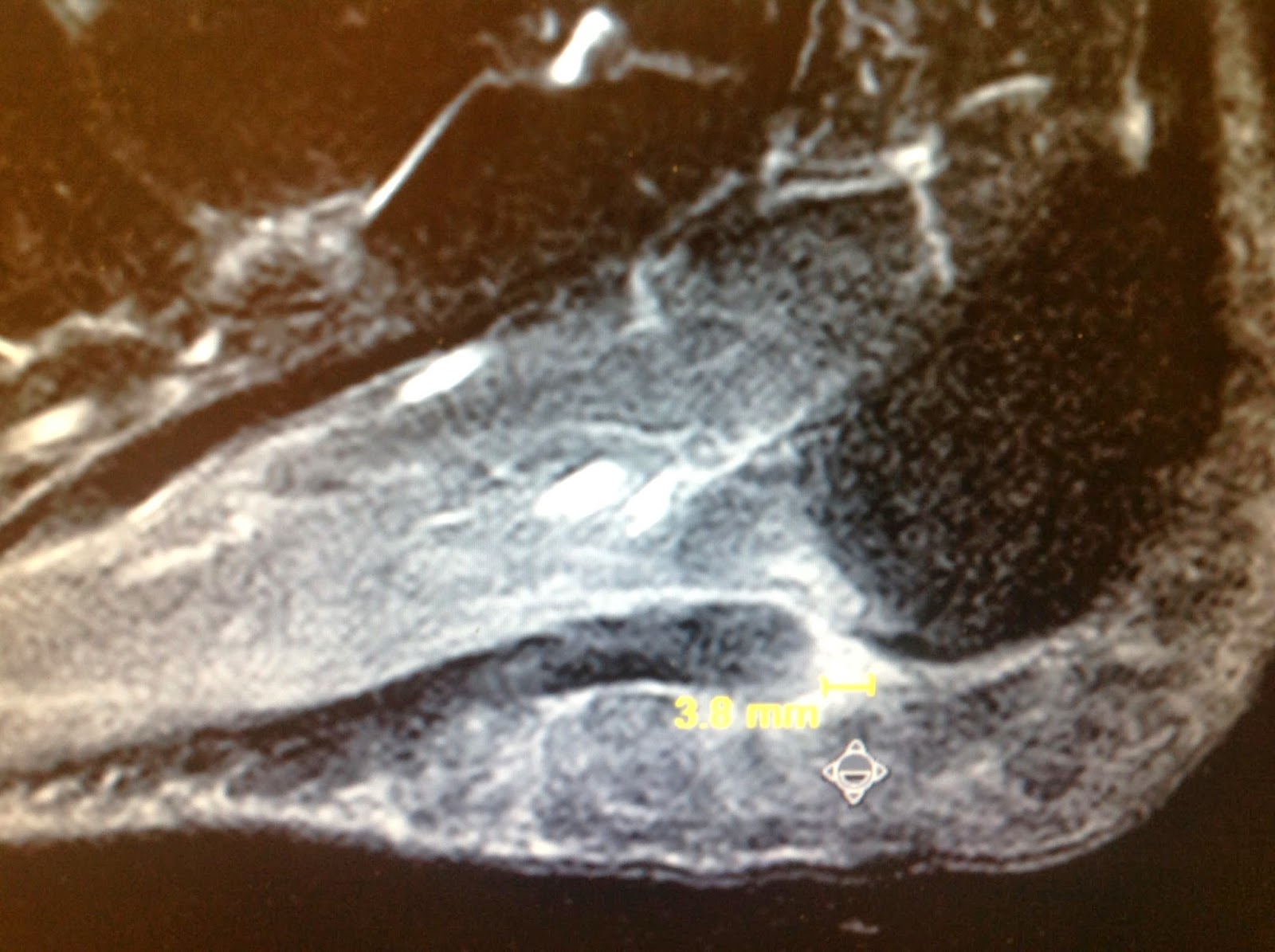
Limb-length discrepancies or differences are conditions that result in limbs that are unequal in length or that exhibit other anomalies. The conditions occur when bones or joints in the arms or legs are abnormal or become damaged. Providers might refer to these conditions as limb-length discrepancies, limb differences or limb-length conditions.  Causes A patient?s legs may be different lengths for a number of reasons, including a broken leg bone may heal in a shorter position, particularly if the injury was severe. In children, broken bones may grow faster for a few years after they heal, resulting in one longer leg. If the break was near the growth center, slower growth may ensue. Children, especially infants, who have a bone infection during a growth spurt may have a greater discrepancy. Inflammation of joints, such as juvenile arthritis during growth, may cause unequal leg length. Compensation for spinal or pelvic scoliosis. Bone diseases such as Ollier disease, neurofibromatosis, or multiple hereditary exostoses. Congenital differences. Symptoms The most common symptom of all forms of LLD is chronic backache. In structural LLD the sufferer may also experience arthritis within the knee and hip are, flank pain, plantar fasciitis and metatarsalgia all on the side that is longer. Functional LLD sufferers will see similar conditions on the shorter side. Diagnosis The only way to decipher between anatomical and functional leg length inequalities (you can have both) is by a physical measurement and series of biomechanical tests. It is actually a simple process and gets to the true cause of some runner?s chronic foot, knee, hip and back pain. After the muscles are tested and the legs are measured it may be necessary to get a special X-ray that measures both of your thighs (Femurs) and legs (Tibias). The X-ray is read by a medical radiologist who provides a report of the actual difference down to the micrometer leaving zero room for error. Once the difference in leg length is known, the solution becomes clear. Non Surgical Treatment The key to treatment of LLD in a child is to predict what the discrepancy is at maturity. If it is predicted to be less than 2 cm., no treatment is needed. Limb length discrepancies of up to 2 or 2.5 cm. can be compensated very well with a lift in the shoe. Beyond 2.5 cm., it becomes increasingly difficult to compensate with a left in the insole. Building up the shoe becomes uncosmetic and cumbersome, and some other way of compensating for the discrepancy becomes necessary. The treatment of LLD is long-term treatment, and involves the physician and patient?s family working together as a team. The family needs to weigh the various options available. If leg lengthening is decided on, the family needs to understand the commitment necessary to see it through. The treatment takes 6 months to a year for completion, and complications can happen. But when it works, the results are gratifying.  how to become tall in one day Surgical Treatment Shortening techniques can be used after skeletal maturity to achieve leg length equality. Shortening can be done in the proximal femur using a blade plate or hip screw, in the mid-diaphysis of the femur using a closed intramedullary (IM) technique, or in the tibia. Shortening is an accurate technique and involves a much shorter convalescence than lengthening techniques. Quadriceps weakness may occur with femoral shortenings, especially if a mid-diaphyseal shortening of greater than 10% is done. If the femoral shortening is done proximally, no significant weakness should result. Tibial shortening can be done, but there may be a residual bulkiness to the leg, and risks of nonunion and compartment syndrome are higher. If a tibial shortening is done, shortening over an IM nail and prophylactic compartment release are recommended. We limit the use of shortenings to 4 to 5 cm leg length inequality in patients who are skeletally mature. Overview
 Heel pain is one of the most common conditions treated by podiatrists. It is often a message from the body that something is in need of medical attention. Pain that occurs right after an injury or early in an illness may play a protective role, often warning us about the damage we have suffered. The greatest incidence of heel pain is seen in middle-aged men and women. It is also seen in those who take part in regular sporting activities and those significantly overweight and on their feet a lot. Heel pain can also occur in children, usually between 8 and 13, as they become increasingly active in sporting activities. Causes Age plays a large role in the development of heel pain, particularly among those over 40. Being active is also a common factor of heel pain. Over time, the elasticity of the tissue in our feet decreases with age, causing us to become prone to damage and also slowing the body's ability to heal damage. Adolescents are also not immune to heel pain. Those who are active in sports are particularly prone to excessively stretching or straining the plantar fascia or Achilles tendon, causing severe heel pain. In most cases, heel pain develops in only one heel. There are many risk factors that lead to heel pain. Abnormal gait and excessive, repetitive stress are common factors in the development of pain and damage. Among the other risk factors involved with the development of heel pain are repetitive exercise or activities, such as long distance running or jumping from activities such as basketball. Obesity. Walking barefoot on hard surfaces. Prolonged standing. Wearing poor fitting shoes, or shoes that do not provide enough support or cushioning. Not stretching properly or at all before and after exercise. Those who are on their feet for long periods of time. Symptoms Pain in the heel can be caused by many things. The commonest cause is plantar fascitis. Other causes include, being overweight, constantly being on your feet, especially on a hard surface like concrete and wearing hard-soled footwear, thinning or weakness of the fat pads of the heel, injury to the bones or padding of the heel, arthritis in the ankle or heel (subtalar) joint, irritation of the nerves on the inner or outer sides of the heel, fracture of the heel bone (calcaneum). Diagnosis Your doctor will perform a physical exam and ask questions about your medical history and symptoms, such as have you had this type of heel pain before? When did your pain begin? Do you have pain upon your first steps in the morning or after your first steps after rest? Is the pain dull and aching or sharp and stabbing? Is it worse after exercise? Is it worse when standing? Did you fall or twist your ankle recently? Are you a runner? If so, how far and how often do you run? Do you walk or stand for long periods of time? What kind of shoes do you wear? Do you have any other symptoms? Your doctor may order a foot x-ray. You may need to see a physical therapist to learn exercises to stretch and strengthen your foot. Your doctor may recommend a night splint to help stretch your foot. Surgery may be recommended in some cases. Non Surgical Treatment Curing posterior heel pain requires calming the inflammation, resting the foot and increasing calf flexibility. Ice therapy and anti-inflammatory medications can be used to reduce the inflammation. Stopping exercises that stress the calf and Achilles is important. This includes walking, running and the use of stair climbers and elliptical machines. Placing a heel lift in each shoe can reduce some of the excess strain on the back of the heel. Stretching exercises to increase calf flexibility are important in curing this problem and preventing its recurrence. Wearing running shoes also provides good foot support and helps with this condition. Sometimes a walking boot is used to immobilize the ankle and let the area completely rest. Physical therapy is sometimes ordered to help reduce the inflammation and pain and to help improve the flexibility of the calf muscles. Occasionally these measures fail to relieve the pain and surgery may be needed. The surgical procedure involves removing bone spurs and repairing any damage to the tendon. Surgical Treatment When a diagnosis of plantar fasciitis is made early, most patients respond to conservative treatment and don?t require surgical intervention. Often, when there is a secondary diagnosis contributing to your pain, such as an entrapped nerve, and you are non-responsive to conservative care, surgery may be considered. Dr. Talarico will discuss all options and which approach would be the most beneficial for your condition. heel pads shoes too big Prevention  It may not be possible to prevent all cases of heel pain. However, there are some easy steps that you can take to avoid injury to the heel and prevent pain. Whenever possible, you should wear shoes that fit properly and support the foot, wear the right shoes for physical activity, stretch your muscles before exercising, pace yourself during physical activity, maintain a healthy diet, rest when you feel tired or when your muscles ache, maintain a healthy weight. Overview
 A neuroma is a painful condition, also referred to as a ?pinched nerve? or a nerve tumor. It is a benign growth of nerve tissue frequently found between the third and fourth toes. It brings on pain, a burning sensation, tingling, or numbness between the toes and in the ball of the foot. The principal symptom associated with a neuroma is pain between the toes while walking. Those suffering from the condition often find relief by stopping their walk, taking off their shoe, and rubbing the affected area. At times, the patient will describe the pain as similar to having a stone in his or her shoe. The vast majority of people who develop neuromas are women. A neuroma is a painful condition, also referred to as a ?pinched nerve? or a nerve tumor. It is a benign growth of nerve tissue frequently found between the third and fourth toes. It brings on pain, a burning sensation, tingling, or numbness between the toes and in the ball of the foot. The principal symptom associated with a neuroma is pain between the toes while walking. Those suffering from the condition often find relief by stopping their walk, taking off their shoe, and rubbing the affected area. At times, the patient will describe the pain as similar to having a stone in his or her shoe. The vast majority of people who develop neuromas are women.Causes Pronation of the foot can cause the metatarsal heads to rotate slightly and pinch the nerve running between the metatarsal heads. This chronic pinching can make the nerve sheath enlarge. As it enlarges it than becomes more squeezed and increasingly troublesome. Tight shoes, shoes with little room for the forefoot, pointy toeboxes can all make this problem more painful. Walking barefoot may also be painful, since the foot may be functioning in an over-pronated position. Symptoms Patients will feel pain that worsens with walking, particularly when walking in shoes with thin soles or high heels. Also, anything that squeezes the metatarsal heads together may aggravate symptoms, such as narrow shoes. A patient may feel the need to remove the shoe and rub the foot to soothe the pain. Diagnosis The most common condition misdiagnosed as Morton's neuroma is metatarsophalangeal (MTP) joint synovitis. When pain occurs in the third interspace, the clinician may misdiagnose the condition as Morton's neuroma instead of MTP synovitis, which may manifest very much like Morton's neuroma. MTP synovitis is distinguished from Morton's neuroma by subtle swelling around the joint, pain localized mainly within the joint, and pain with forced toe flexion. Palpation of the MTP joint is performed best with a pinching maneuver from the dorsal and plantar aspects of the joint to elicit tenderness of the joint. Other conditions often misdiagnosed as Morton's neuroma include the following. Stress fracture of the neck of the metatarsal. Rheumatoid arthritis and other systemic arthritic conditions. Hammertoe. Metatarsalgia (ie, plantar tenderness over the metatarsal head) Less common conditions that have overlapping symptoms with Morton's neuroma include the following. Neoplasms. Metatarsal head osteonecrosis. Freiburg. steochondrosis. Ganglion cysts. Intermetatarsal bursal fluid collections. True neuromas. Non Surgical Treatment Most patients' symptoms subside when they change footwear to a wide soft shoe with a metatarsal support inside to relieve the pressure on the involved area. If this treatment fails, a cortisone injection into the nerve is occasionally helpful.  Surgical Treatment Patients are commonly offered surgery known as neurectomy, which involves removing the affected piece of nerve tissue. Postoperative scar tissue formation (known as stump neuroma) can occur in approximately 20%-30% of cases, causing a return of neuroma symptoms. Neurectomy can be performed using one of two general methods. Making the incision from the dorsal side (the top of the foot) is the more common method but requires cutting the deep transverse metatarsal ligament that connects the 3rd and 4th metatarsals in order to access the nerve beneath it. This results in exaggerated postoperative splaying of the 3rd and 4th digits (toes) due to the loss of the supporting ligamentous structure. This has aesthetic concerns for some patients and possible though unquantified long-term implications for foot structure and health. Alternatively, making the incision from the ventral side (the sole of the foot) allows more direct access to the affected nerve without cutting other structures. However, this approach requires a greater post-operative recovery time where the patient must avoid weight bearing on the affected foot because the ventral aspect of the foot is more highly enervated and impacted by pressure when standing. It also has an increased risk that scar tissue will form in a location that causes ongoing pain. There are two different types of leg length discrepancies, congenital and acquired. Congenital means that you are born with it. One leg is structurally shorter in comparison to the other. Through developmental periods of aging, the human brain picks up on the walking pattern and identifies some variation. The body typically adapts by dipping one shoulder to the "short" side. A difference of less than a quarter inch is not very excessive, does not need Shoe Lifts to compensate and generally doesn't have a serious effect over a lifetime.
 Leg length inequality goes mainly undiagnosed on a daily basis, however this problem is simply fixed, and can eliminate a number of instances of back discomfort. Treatment for leg length inequality usually involves Shoe Lifts. These are very reasonably priced, often being below twenty dollars, compared to a custom orthotic of $200 if not more. When the amount of leg length inequality begins to exceed half an inch, a whole sole lift is generally the better choice than a heel lift. This prevents the foot from being unnecessarily stressed in an abnormal position. Chronic back pain is the most common ailment affecting people today. Around 80 million men and women experience back pain at some stage in their life. It's a problem which costs companies huge amounts of money annually because of time lost and production. Innovative and improved treatment solutions are constantly sought after in the hope of minimizing the economical influence this issue causes.  People from all corners of the world suffer the pain of foot ache due to leg length discrepancy. In these situations Shoe Lifts are usually of very helpful. The lifts are capable of reducing any discomfort in the feet. Shoe Lifts are recommended by countless experienced orthopaedic doctors. In order to support the human body in a balanced fashion, the feet have got a crucial job to play. Despite that, it is sometimes the most neglected area of the body. Many people have flat-feet meaning there is unequal force placed on the feet. This will cause other areas of the body like knees, ankles and backs to be impacted too. Shoe Lifts ensure that ideal posture and balance are restored.  Overview OverviewThe hammertoes condition is usually irreversible, but often its progression can be slowed or halted. You should visit a Podiatrist if the toe becomes painful and you have difficulty walking. A Podiatrist will be able to provide advice and treatment including padding the bony top-part of your hammertoe to relieve pain or to tape your toes as a way to change their position. Podiatrists have an important role to play in preventing and managing foot problems. Prompt action is important. Problems which are left without assessment or treatment may result in major health risks. Causes Your toe contains two joints that allow it to bend at the middle and bottom. A hammertoe occurs when the middle joint becomes dislocated. Common causes of this joint dislocation include a toe injury, arthritis, a high foot arch, wearing shoes that don?t fit properly, tightened ligaments or tendons in the foot, pressure from a bunion (when your big toe points inward toward your second toe) Spinal cord or peripheral nerve damage may cause all of your toes to curl downward.  Symptoms SymptomsThe most obvious symptom of hammer, claw or mallet toe is the abnormal toe position. This is usually combined with pain: the abnormal foot position leads to excessive friction on the toe as it rubs against any footwear which can be extremely painful. Corns & Calluses: repeated friction can result in the formation of a foot corn or callus on top of the toes. Stiffness, the joints become increasingly stiff. In the early stages, the toes can usually be straightened out passively using your hands, but if allowed to progress, the stiffness may be permanent. Diagnosis First push up on the bottom of the metatarsal head associated with the affected toe and see if the toe straightens out. If it does, then an orthotic could correct the problem, usually with a metatarsal pad. If the toe does not straighten out when the metatarsal head is pushed up, then that indicates that contracture in the capsule and ligaments (capsule contracts because the joint was in the wrong position for too long) of the MTP joint has set in and surgery is required. Orthotics are generally required post-surgically. Non Surgical Treatment If the toes are still mobile enough that they are able to stretch out and lay flat, the doctor will likely suggest a change of footwear. In addition, she may choose to treat the pain that may result from the condition. The doctor may prescribe pads to ease the pain of any corns and calluses, and medications ranging from ibuprofen to steroid injections for the inflammation and pain. Other options for non-surgical treatments include orthotic devices to help with the tendon and muscle imbalance or splinting to help realign the toe. Splinting devices come in a variety of shapes and sizes but the purpose of each is Hammer toes the same: to stretch the muscles and tendon and flatten the joint to remove the pain and pressure that comes from corns. Surgical Treatment In advanced cases in which the toe has become stiff and permanently bent, the toe can be straightened with surgery. One type of surgery involves removing a small section of the toe bone to allow the toe to lie flat. Surgery for hammertoe usually is classified as a cosmetic procedure. Cosmetic foot surgeries sometimes cause complications such as pain or numbness, so it?s better to treat the problem with a shoe that fits properly.  Overview OverviewHammer Toe is a condition which causes one or more of the smaller toes to become bent upwards. The toe can be straightened but if ignored may become a permanent deformity. Each of the 4 smaller toes consist of 3 bones called phalanges, forming two interphalangeal joints. The toe bends at the proximal or first interphalangeal joint. Initially it can be straightened, but if left untreated, this can become a permanent deformity. Causes Hammertoe is caused when muscles fail to work in a balanced manner and the toe joints bend to form the hammertoe shape. If they remain in this position, the muscles and tendons supporting them tighten and stay that way. Causes of hammertoe can include squeezing into a too-small or ill-fitting shoe or wearing high heels that jam your toes into a tight toe box. An injury such as badly stubbing your toe. Arthritis. Nerve and muscle damage from diseases such as diabetes,  Symptoms If you have any of these symptoms, do not assume it is due to hammer toe. Talk to your doctor about symptoms such as a toe that curls down, corns on the top of a toe, calluses on the sole of the foot or bottom of the toe, pain in the middle joint of a toe, discomfort on the top of a toe, difficulty finding any shoes that fit comfortably, cramping in a toe, and sometimes also the foot and leg, difficult or painful motion of a toe joint, pain in the ball of the foot or at the base of a toe. Diagnosis The earlier a hammertoe is diagnosed, the better the prognosis and treatment options. Your doctor will be able to diagnose your hammertoe with a simple examination of the foot and your footwear. He or she may take an x-ray to check the severity of the condition. You may also be asked about your symptoms, your normal daily activities, and your medical and family history. Non Surgical Treatment You can usually use over-the-counter cushions, pads, or medications to treat bunions and corns. However, if they are painful or if they have caused your toes to become deformed, your doctor may opt to surgically remove them. If you have blisters on your toes, do not pop them. Popping blisters can cause pain and infection. Use over-the-counter creams and cushions to relieve pain and keep blisters from rubbing against the inside of your shoes. Gently stretching your toes can also help relieve pain and reposition the affected toe. Surgical Treatment Bone-mending procedures realign the contracted toe by removing the entire deviated small joints of the toe (again, not at the ball of the foot). This allows for the buckled joint to be positioned flat and the bone ends to mend together. Often surgical hardware (fixation) is necessary to keep the bones steady during healing. Hardware options can involve a buried implant inside the toe, or a temporary wire that is removed at a later date. Medical terminology for this procedure is called a proximal interphalangeal joint arthrodesis (fusion), or a distal interphalangeal joint arthrodesis (fusion), with the former being performed in a high majority of cases.
Overview
 Bunions are bony bumps that form on the side of the big toe joint or baby toe joint (these are commonly called ?bunionettes? or ?tailor?s bunions?). Bunions most commonly result from one or more of the following factors: genetics, faulty foot mechanics like over-pronation, and/or long periods of time spent in improperly fitting footwear. Bunions can start with audible clicking (called ?crepitus?) and/or stiffness in the affected joint which indicates that the joint surfaces are rubbing together improperly. This may progress to include inflammation, degeneration of the surfaces of the joint, deformity (including bone growth at the joint line and displacement of the toe) and ultimately, loss of range of motion in the joint. Bunions are bony bumps that form on the side of the big toe joint or baby toe joint (these are commonly called ?bunionettes? or ?tailor?s bunions?). Bunions most commonly result from one or more of the following factors: genetics, faulty foot mechanics like over-pronation, and/or long periods of time spent in improperly fitting footwear. Bunions can start with audible clicking (called ?crepitus?) and/or stiffness in the affected joint which indicates that the joint surfaces are rubbing together improperly. This may progress to include inflammation, degeneration of the surfaces of the joint, deformity (including bone growth at the joint line and displacement of the toe) and ultimately, loss of range of motion in the joint.Causes High heels can exacerbate a potential bunion problem because they tip the body?s weight forward, forcing the toes into the front of the shoe. This may help to explain why bunions are 10 times more common in women than in men. People in occupations such as teaching and nursing, that involve a lot of standing and walking, are susceptible to bunions. Ballet dancers, whose feet suffer severe repetitive stress, are also amongst those who experience bunions. Women can sometimes develop bunions and other foot problems during pregnancy because hormonal changes loosen the ligaments and flatten the feet. Bunions are also associated with arthritis, which damages the cartilage within the joint. Symptoms Bunions may cause no pain at first. But as the big toe begins to turn in towards the other toes, people with bunions usually experience redness, pain, swelling, and tenderness in the area around the joint. Pressure inside the joint or from footwear pressing against the bunion may also cause discomfort. As the affected toe curves closer to the other toes on the foot, these toes can become painful as well. Complications of bunions include corns, calluses, hammer toe, and ingrown toenails. Other complications include irritation of the nerves surrounding the bunion area. Excess rubbing of the bunion against the footwear may lead to changes in the skin, resulting in corns or calluses. Hammer toe is a deformity of the toe immediately next to the big toe. A hammer toe is slightly raised and points upwards from the base and downwards at the end of the toe. Ingrown toenails can result from increased pressure from the big toe on the other toes. There may also be a decrease in the amount a person can move the joint affected by the bunion. Irritation of the nerves will feel like burning or decreased sensation. Diagnosis Your doctor will ask questions about your past health and carefully examine your toe and joint. Some of the questions might be: When did the bunions start? What activities or shoes make your bunions worse? Do any other joints hurt? The doctor will examine your toe and joint and check their range of motion. This is done while you are sitting and while you are standing so that the doctor can see the toe and joint at rest and while bearing weight. X-rays are often used to check for bone problems or to rule out other causes of pain and swelling. Other tests, such as blood tests or arthrocentesis (removal of fluid from a joint for testing), are sometimes done to check for other problems that can cause joint pain and swelling. These problems might include gout , rheumatoid arthritis , or joint infection. Non Surgical Treatment Conservative Treatment. Apply a commercial, nonmedicated bunion pad around the bony prominence. Wear shoes with a wide and deep toe box. If your bunion becomes inflamed and painful, apply ice packs several times a day to reduce swelling. Avoid high-heeled shoes over two inches tall. See your podiatric physician if pain persists. Orthotics. Shoe inserts may be useful in controlling foot function and may reduce symptoms and prevent worsening of the deformity. Padding & Taping. Often the first step in a treatment plan, padding the bunion minimizes pain and allows the patient to continue a normal, active life. Taping helps keep the foot in a normal position, thus reducing stress and pain. Medication. Anti-inflammatory drugs and cortisone injections are often prescribed to ease the acute pain and inflammations caused by joint deformities. Physical Therapy. Often used to provide relief of the inflammation and from bunion pain. Ultrasound therapy is a popular technique for treating bunions and their associated soft tissue involvement.  Surgical Treatment When these above measures no longer help to relieve the pain in the big toe, surgery to correct the bunion deformity is considered. Numerous surgical procedures have been recommended for bunions. What is most critical is that the type of deformity is carefully evaluated, because one bunion surgery cannot be used for all types of bunions. If the big toe joint is rotated out of place, the joint must be rotated back in place for the procedure to work. Conversely, a bunion can occur with the big toe still ?in place.? If surgery is considered, the bunion must be corrected with the toe joint left in its current position. In other words, one type of bunion repair does not work for everyone. In all types of bunion repairs, ligaments and tendons (soft tissues) around the big toe joint are reconstructed, to allow the toe to be straightened. Most bunion procedures also require cutting the metatarsal bone, which is then fixed with metal screws to hold the bone in position until it heals. It usually takes 2 to 4 months to fully recover from bunion surgery, which is why it is always the last course of treatment. Prevention Bunions often become painful if they are allowed to progress. But not all bunions progress. Many bunion problems can be managed without surgery. In general, bunions that are not painful do not need surgical correction. For this reason, orthopaedic surgeons do not recommend ?preventive? surgery for bunions that do not hurt, with proper preventive care, they may never become a problem. |
